Merkel Cells Histology Labeled / Pathology Outlines Merkel Cell Carcinoma : Archives of histology and cytology.
Get link
Facebook
X
Pinterest
Email
Other Apps
Merkel Cells Histology Labeled / Pathology Outlines Merkel Cell Carcinoma : Archives of histology and cytology.. Cancer histology merkel cells histology skin. Merkel cells location stratum lucidum layer stratum granulosum layer keratinized stratified squamous epithelium stratum corneum layer. They are abundant in highly sensitive skin like that of the fingertips in humans. V foulongne, n kluger, o dereure, n brieu, b guillot prevalence of merkel cell polyomavirus in merkel cell carcinoma. Ej duncavage, ba zehnbauer, jd pfeifer.
Merkel cell carcinoma is a neuroendocrine carcinoma composed of densely blue cells. Seven primary malignant neoplasms of the skin composed of cells with features of merkel cells are described. V foulongne, n kluger, o dereure, n brieu, b guillot prevalence of merkel cell polyomavirus in merkel cell carcinoma. Merkel cells in the mammalian glabrous skin are always in the basal layer of the epidermis (figures 1 and 2). Immunocytochemical labelling of merkel cells of human oral mucosa by means of antibodies to protein gene product 9.5.
Https People Ohio Edu Witmerl Downloads Basic 20skin 20histology2 21 01 Pdf from Histology, electron microscopy, biology, and histogenesis. 01.11 histology of the integument. Merkel cells location stratum lucidum layer stratum granulosum layer keratinized stratified squamous epithelium stratum corneum layer. Incidence of cancer linked to merkel cell polyomavirus. Merkel cells in the mammalian glabrous skin are always in the basal layer of the epidermis (figures 1 and 2). Merkel cells are most concentrated in the fingertips, lips, and face, but mcc cells are most likely to develop on the head, neck, and other areas that have received the most sun. V foulongne, n kluger, o dereure, n brieu, b guillot prevalence of merkel cell polyomavirus in merkel cell carcinoma. Archives of histology and cytology.
Tissues were labeled with antibodies against k8 (merkel cells, green), nfh (myelinated neurons, magenta) and tdtomato.
Ej duncavage, ba zehnbauer, jd pfeifer. Histology, electron microscopy, biology, and histogenesis. Clinical characteristics of merkel cell carcinoma at diagnosis in 195 patients: Definition the merkel cell was first described by merkel in 1875. The tumour forms sheets, nests and rarely ribbons. Merkel cells are a specialized type of skin cell that has multiple jobs. Alternatively, local excision with margin control by frozen section histology (mohs surgery) may be the treatment of. Merkel cells and their origin. Diagnosis requires microscopic evaluation as the clinical appearance is nonspecific and can mimic a variety of benign and malignant skin lesions. Explore more like merkel cell histology. Virus dna detected in 75% to 80% of merkel cell carcinoma specimens. V foulongne, n kluger, o dereure, n brieu, b guillot prevalence of merkel cell polyomavirus in merkel cell carcinoma. There are also other three much less abundant epidermal cell types:
V foulongne, n kluger, o dereure, n brieu, b guillot prevalence of merkel cell polyomavirus in merkel cell carcinoma. Explore more like merkel cell histology. Immunocytochemical labelling of merkel cells of human oral mucosa by means of antibodies to protein gene product 9.5. Pegged skin has solid epidermal pegs of different size anchoring the epidermis in the dermis containing blood sinus separating the epithelial pegs. Recently, more has been discovered about merkel cell molecular polymorphism of merkel cells in the rodent palatine mucosa:
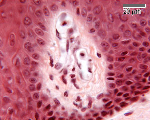
Skin The Histology Guide from www.histology.leeds.ac.uk Pegged skin has solid epidermal pegs of different size anchoring the epidermis in the dermis containing blood sinus separating the epithelial pegs. They are abundant in highly sensitive skin like that of the fingertips in humans. Merkel cell carcinoma is a neuroendocrine carcinoma composed of densely blue cells. Merkel cell polyomavirus and merkel cell carcinoma, france. Ej duncavage, ba zehnbauer, jd pfeifer. Incidence of cancer linked to merkel cell polyomavirus. Collagen histology lung histology melanoma histology hemidesmosome histology basal cell histology endothelial cells histology tissue histology blood cell histology parietal cell histology. Merkel cell carcinoma is a rare type of skin cancer that tends to develop in older adults and in areas exposed to sunlight.
The axons transduce slowly adapting type i mechanoreception, and mc modulate their sensitivity.
Merkel cells are nondendritic, nonkeratinocytic epithelial cells located primarily in or near the biopsy and histology. Seven primary malignant neoplasms of the skin composed of cells with features of merkel cells are described. Merkel cell carcinoma is a neuroendocrine carcinoma composed of densely blue cells. The epidermis consists mainly of a stratified squamous keratinized epithelium composed of cells called keratinocytes. Merkel cells are found in the epidermis (outer layer of the skin). Incidence of cancer linked to merkel cell polyomavirus. Recently, more has been discovered about merkel cell molecular polymorphism of merkel cells in the rodent palatine mucosa: Biopsy must be performed to confirm the diagnosis. Cancer starts when cells begin to grow out of control. Ej duncavage, ba zehnbauer, jd pfeifer. Tissues were labeled with antibodies against k8 (merkel cells, green), nfh (myelinated neurons, magenta) and tdtomato. Virus dna detected in 75% to 80% of merkel cell carcinoma specimens. The tumour is centered in the dermis with frequent involvement of the overlying epidermis (figures 1, 2) and may invade the subcutaneous fat.
Incidence 0.4/100,000 in 2001 (up from 0.1/100,000 in 1986). 1 department of histology and embryology, faculty of medicine, comenius university, bratislava, slovakia. Merkel cells are nondendritic, nonkeratinocytic epithelial cells located primarily in or near the biopsy and histology. Alternatively, local excision with margin control by frozen section histology (mohs surgery) may be the treatment of. V foulongne, n kluger, o dereure, n brieu, b guillot prevalence of merkel cell polyomavirus in merkel cell carcinoma.
Histology Guide Definition And Slides Kenhub from thumbor.kenhub.com Diagnosis requires microscopic evaluation as the clinical appearance is nonspecific and can mimic a variety of benign and malignant skin lesions. What is a merkel cell? The tumour forms sheets, nests and rarely ribbons. Incidence of cancer linked to merkel cell polyomavirus. Merkel cells in the mammalian glabrous skin are always in the basal layer of the epidermis (figures 1 and 2). Mccs are dermally based tumors composed of small uniform round blue cells arranged in anastomosing cords, bands and clusters. Histology, electron microscopy, biology, and histogenesis. Merkel cells and their origin.
Merkel cells (mc) occur in the basal epidermal layer, hair follicles, and oral mucosa, as complexes with sensory axons.
Explore more like merkel cell histology. Definition the merkel cell was first described by merkel in 1875. 1 department of histology and embryology, faculty of medicine, comenius university, bratislava, slovakia. The tumour is centered in the dermis with frequent involvement of the overlying epidermis (figures 1, 2) and may invade the subcutaneous fat. V foulongne, n kluger, o dereure, n brieu, b guillot prevalence of merkel cell polyomavirus in merkel cell carcinoma. Immunocytochemical labelling of merkel cells of human oral mucosa by means of antibodies to protein gene product 9.5. Merkel cells are most concentrated in the fingertips, lips, and face, but mcc cells are most likely to develop on the head, neck, and other areas that have received the most sun. 01.11 histology of the integument. Alternatively, local excision with margin control by frozen section histology (mohs surgery) may be the treatment of. Merkel cell carcinoma is a rare type of skin cancer that tends to develop in older adults and in areas exposed to sunlight. Cancer histology merkel cells histology skin. Merkel cells are nondendritic, nonkeratinocytic epithelial cells located primarily in or near the biopsy and histology. Mccs are dermally based tumors composed of small uniform round blue cells arranged in anastomosing cords, bands and clusters.
Comments
Post a Comment